| 1 |
|
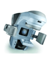
Linear accelerator therapy |
CLINAC IX |
Radiation Oncology Clinic |
With the introduction of HELAX (a dedicated treatment planning system for IMRT computers), which uses the most accurate algorithms developed so far, and the CT-Simulator (AcQ-Sim) that constructs 3-D images of the internal body, the hospital now supports a complete IMRT implementation. Intensity Modulated Radiation Therapy is already being used for many indications including head and neck cancer and prostate cancer while the usage will soon spread to the breast, esophagus and the uterus. |
| 2 |
|
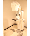
Breast X-ray digital imaging equipment |
|
Health Promotion Center |
This method creates an image of X-rays using digital detectors which shows the smallest tissues in the breast with a resolution higher than most films. Due to the small breast size and dense breast tissues of Korean women, it was difficult to differentiate lesions from normal tissues; however, with the high resolution digital breast mammography, the contrast is increased between normal tissue and lesions for easy differentiation. |
| 3 |
|
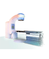
Linear accelerator (CT-SIMULATOR) |
LIGHT-SPEED RT/GE |
Radiation Oncology Clinic |
With the simulation for radiotherapy treatment tomography, 1mm sections can be seen in detail in 3-D and the extent of movement of the organs according to breathing can be captured with 50% less radiation exposure compared to existing methods. |
| 4 |
|
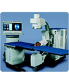
Extracorporeal Shock Wave Crusher |
Compact-Delta |
Urology outpatient |
This is a procedure for removing kidney stones with shock waves which are administered from the outside of the body. The shock waves break the kidney stones into small pieces which are then naturally eliminated in urine.
It was found that 90% of urinary stones can be removed by extracorporeal shock wave lithotripsy and the success rate was 90%. The procedure can be administered without anesthesia or incision or sequelae and many patients prefer this procedure. |
| 5 |
|
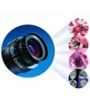
OPERATION MICROSCOPE |
OPMI Neuro in NC4 Fl |
Operating room |
The head part moves freely so that the observation can easily be made from different angles. The vertical movement and mounting of foot switch is an X-Y translator which is useful in micro-surgery. |
| 6 |
|
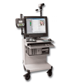
EEG/Video monitoring sys(S3) |
Beehive Horizon |
Nervous system laboratory |
A continuous video EEG device from channels 2 to 128, basic functions include audio, recording and auto seizure detecting program with PSG. |
| 7 |
|
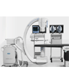
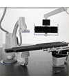
Angiographic X-ray unit / cardiovascular |
AXIOM Artis dFC |
Cardiovascular imaging room |
In addition to coronary artery disease (angina and myocardial infarction), all types of heart diseases including valvular heart disease, arrhythmia, hypertension, hyperlipidemia and myocardial disease are diagnosed and treated with the latest echocardiography and cardiovascular imaging system. In the present when the intervention procedures are becoming more complex, it provides an excellent high-resolution image while reducing the exposure to x-ray without compromising resolution. Also, the user can change/set the repeated parameters manually when the optimal image resolution is known. |
| 8 |
|
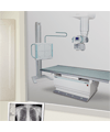
Digital X-Ray system - 1Detector(DRS-1000) |
DRS(CHEST DR FULL SY) |
Radiology Clinic |
DRS is a digital filming system compatible with DICOM and boasts perfect compatibility along with filmless environment. Specifically, it is equipped with the latest technology and the worklist function for HIS/RIS integration so that accurate diagnosis can be made in a short period of time with the efficient controls and high-resolution image. |
| 9 |
|
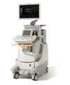
Ultrasound imaging device (IU22) |
IU22 |
Radiology Clinic |
This is the latest ultrasound imaging diagnostic device which uses harmless ultrasound to show the tissues on the monitor. This device is a state-of-the-art device with color output that can diagnose vascular health just by spectral doppler.
This device can be used to diagnose everything on the body - PureWave Crystal Technology can be used for heart examinations as well as in general imaging such as thyroid problems, breast cancer, abdomen, gynecology and urinary system. |
| 10 |
|
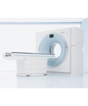
CT System |
SOMATOM Sensation 64 |
Radiology Clinic |
While the existing CT device captures 1 image per rotation, 64-channel CT can capture 64 images with 64 times the speed. Therefore, it can diagnose the location and the status with more accuracy.
The all-in-one examinations for coronary artery disease, myocardium and cardiac function scans are possible with the newly introduced 64-channel CT. As it can show the general status of the internal organs without being inserted into the body, using this has minimized the pain for patients as it can give a comprehensive evaluation on the narrowing of the blood vessels, malformations, bronchiectasis, incision of the blood vessels increasing, pulmonary artery embolism and is useful in diagnosing vascular diseases as well as treating strokes. |
| 11 |
|
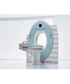
CT System |
SOMATOM Sensation 128 |
Radiology Clinic |
It is faster than the existing 64-channel for cardiac recording and the radiation amount has been drastically reduced to half that of MDCT. Specifically, this is good news for cardiovascular patients who had to wait or for whom the recording was not possible at all because of high-resolution and faster device speed. Patients whose heart rates were too high and required injections to slow it down can now quickly go through the examination process. |
| 12 |
|
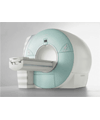
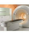
MRI SYSTEM |
MAGNETOM Avanto |
Radiology Clinic |
Accurate diagnosis and treatment is possible with the selection of SQ engine and 18 detection channels that can give detailed images in short time Examination time is dramatically shortened - whole body check up takes place in 12 minutes, back check up in 20 minutes Shortened examination time is very practical for people with reduced mobility or for children and even those who feel claustrophobic. |
| 13 |
|
ANGIO X-RAY SYSTEM /SINGLE |
AXIOM Artis dFA |
Radiology Clinic |
The heart, aorta, brain, blood vessels, abdominal aorta and its branches. This device allows for the X-ray imaging of blood vessels in order to scan for any defects in the blood system. |
| 14 |
|
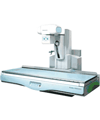
Digital Remote R/F X-ray system(Sonialvision 100) |
SONIALVISION 100 |
Radiology Clinic |
This device can be used with all types of fluoroscopy including gastrointestinal/colorectal scan, non-vascular interventional procedures, vascular imaging and tomography. This method does not require the use of IP cassette during a scan as it is already equipped with a flat panel detector that uses direct conversion method of a-Se devices. Also, there is no damage to images as in 1.1 and so high quality images can be obtained with a small amount of X-ray. Also, the device can be controlled with a remote in order to prevent radiation; adjustable height of the table allows for easy access for those with limited mobility. |
| 15 |
|
Sample pretreatment automation equipment |
cobas 8000 |
Laboratory Medicine |
The best advantage is that the 8400 test per hour is the fastest examination time. Specimens can be mounted during the inspection and the track as well as MSB (Module Sample Buffer) can increase the efficacy of the examination room. In addition, speeding up the examination process increases the convenience for patients as they wait for a shorter time for the results. |
| 16 |
|
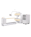
Gamma Camera |
FORTE |
Nuclear Medicine |
A camera that records the structure of the internal organ using radioactive isotopes (hereafter RI) When organ-friendly substances marked with RI are injected into a patient, the substances will disperse in certain organs with a unique pattern. The γ rays are detected from outside the body and then plotted on a XY axis which is useful as an organ scan or a cancer test. |
| 17 |
|
Positron Emission Tomography |
PET-CT/GEMINI 16 |
Nuclear Medicine |
With one examination, tumors (early diagnosis of cancer / differential diagnosis of benign and malignant tumors / cancer staging / recurrence of cancer found / malignant cancer assessment), neuropsychology disorders (diagnosis of dementia and the causes of epilepsy patients / preoperative) and cardiovascular disease (myocardial viability assessment) can be diagnosed. Early detection of cancer is also possible which is difficult with CT or MRI. Also, with just one examination, cancer can be diagnosed with differentiation between malignant/benign tumors possible. The exam is relatively safe with low amounts of radiation. |


